25.09.2024 Freight of small vesicles offers new treatment approach for pulmonary fibrosis
Now published in JCI Insight
A healthy lung can recover from minor injuries caused by viruses or pollutants. However, this regenerative capacity is impaired in patients with pulmonary fibrosis. A research team at the Philipps University of Marburg has now discovered that in pulmonary fibrosis, small vesicles transport a protein that is responsible for the impaired regenerative capacity of the lung. The protein represents a possible target for new therapies and could serve as a biomarker to better detect pulmonary fibrosis.
Idiopathic pulmonary fibrosis is a fatal lung disease that is incurable to date. Typical symptoms of the disease are a decline in lung function and an increase in scarring of the lungs by connective tissue. However, little is known about how pulmonary fibrosis develops.

Prof. Mareike Lehmann and her team at the Philipps-Universität Marburg already knew from their previous work that small cell vesicles, known as extracellular vesicles, are found in increased numbers in the lungs of patients with pulmonary fibrosis. They have now identified the proteins that these vesicles transport and were able to show that the vesicles' cargo increases damage to the lung tissue.
Extracellular vesicles (EVs) are small bubbles that can be released and reabsorbed by almost any cell. They contain molecules such as proteins, lipids and RNA. Their main function is to transport information between cells via their contents.
Reprogramming promotes scarring of lung tissue
Together with researchers from the University of Pittsburgh, the University of Burgundy and the Helmholtz Zentrum München, the Marburg scientists discovered that the content of the vesicles of people with pulmonary fibrosis differs significantly from that of healthy individuals. In particular, a specific protein in the vesicles contributes significantly to the progression of the lung disease by reprogramming the epithelial cells of the lungs.
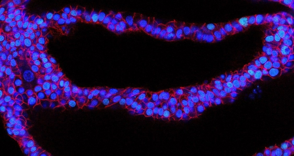
Epithelial cells form the outer protective layer of the lungs and act as a barrier against environmental influences. They can also replace cells and alveolar tissue when damage occurs. The reprogramming means that they can no longer perform this important repair function. Small epithelial injuries can therefore no longer heal, leading to a vicious circle: instead of functional replacement lung tissue, connective tissue cells now form connective tissue, which further exacerbates the disease.
The research team was able to confirm their results at several levels, for example in organoid models, i.e. miniaturized versions of human lungs grown in the laboratory, and in lung tissue and connective tissue cells from patients, which underlines the medical significance.
Targeted intervention in the course of the disease
If the vesicles lacked the protein, the lungs did not scar. The researchers were able to demonstrate this in the mouse model. This provides a new starting point for therapies: “If we could succeed in blocking the protein, it would be possible for the first time to intervene in the course of the disease in a targeted manner,” says Lehmann, who is also a member of the German Center for Lung Research (DZL). Currently available medications can only slow down pulmonary fibrosis, but not stop it. In addition, the disease is difficult to diagnose. If doctors detect a decline in lung function, imaging techniques and tissue samples are needed to confirm the diagnosis. “It would be a significant advance if we could detect the protein in bodily fluids, for example in a lung lavage,” says the Marburg lung researcher.
In addition to targeting the protein, the scientists next want to find out at what stage of the disease the protein appears and how it reacts to the currently available medications.
Original publication: Burgy O, Mayr CH, Schenesse D, et al. Fibroblast-derived extracellular vesicles contain SFRP1 and mediate pulmonary fibrosis. JCI Insight. 2024;9(18): e168889. Published 2024 Aug 15. doi: 10.1172/jci.insight.168889